
Our research process therefore consists of individual steps. The exact procedure depends on the research question and the type of sources involved. Material sources – such as paintings – are examined using methods different from those used for written sources or image sources.
Written sources are evaluated using historical-critical methods. This means that, for example, when reading, one always asks oneself who the author was, and with what aim and in what context the information was recorded by them. This makes it easier to assess what the sources can in actuality contribute to answering the research question.
Material sources such as paintings also contain a great deal of important information about Baumeister's painting technique. We use various methods to decipher this information: Through careful observation, we can find out how he processed his materials and what properties they probably had when he first applied them to his canvases. All selected paintings are therefore first examined with the naked eye and with a stereo microscope, and then photographed.

Willi Baumeister: Discarded painting, ca. 1931, 42 x 28.55 cm, Willi Baumeister Stiftung Stuttgart, Inv. No. V_005.
Photo: Roland Lenz
With the help of a microscope, initial important findings about the material structure of the painting surface and its layer sequence can be obtained. Observations about its condition – whether damage or noticeable changes are evident – are also noted.

Enlarging the image with an optical microscope makes it much easier to see the layers of a painting than with the naked eye. Photo: Elia Schmid
Under magnification, the matte surface of a black paint may reveal traces of application with a very fine brush, for example.

Microscope image of black paint in visible light. Photo: Ulrike Palm
Even in visible light, researchers can make useful observations. However, some things cannot be detected by the naked eye or without the help of imaging devices. This applies, for example, to thin and transparent coatings on the surface of the painting, and to drawings that were later covered with paint. Various examination techniques using different kinds of radiation allow us to make such concealed layers visible without even touching the painting. If during the examination not only visible light, but also other adjacent wavelength ranges such as ultraviolet radiation (UV) or infrared radiation (IR) are used, the paintings reveal further information.

Classification of visible light in the electromagnetic spectrum. The light visible to humans (represented by the black bar labeled with the word “sichtbar”) covers only a very narrow wavelength range. © FWU Institute for Film and Image (CC-BY-NC 4.0).
Available online and reproduced unchanged: https://www.leifphysik.de/optik/elektromagnetisches-spektrum/grundwissen/sichtbares-Licht
Under UV radiation, some materials in coatings or varnishes that are otherwise barely visible develop a pronounced UV fluorescence – also called UV‑induced visible luminescence – and thus can be more easily observed.
In visible light, the surface of the black paint appears very matte, and no coating is visible. Under UV radiation, however, a light blue fluorescent layer on the black paint that was not previously visible in visible light can be observed.
Detail of fragment V_005 in visible light under the stereo microscope.
Photo: Ulrike Palm

Same detail under UV radiation:
On top of the black layer, a blue fluorescent layer becomes visible.
Photo: Ulrike Palm
In the case of Baumeister, we often see such thin, light blue fluorescent layers on the surface, but their exact composition remains to be clarified through further investigation.
Infrared radiation penetrates the surface of the painting and can thus reveal deeper layers of paint, or drawings that are no longer visible to the naked eye because they were covered up later by subsequent paint layers. A special camera is used to record the reflected IR rays, producing a black-and-white image, which is why this method is called infrared reflectography (IRR).

Detail in visible light from Willi Baumeister: Discarded painting, ca. 1931, 42 x 28.55 cm, Willi Baumeister Stiftung Stuttgart, Inv. No. V_005. With the naked eye, pencil lines are visible only in a few places at the edges of the painting.
Photo: Roland Lenz

In infrared reflectography, additional drawing lines become visible that are hidden beneath the layers of paint.
Photo: Roland Lenz
All observations are documented in detail in writing and photographically, and they form an important basis for the subsequent material analyses: Even if the exact materials are not yet known, one can already get an initial overview of the layer structure and the spatial distribution of different materials in the image area. This is important for the selection of suitable – that is, as representative as possible – locations for subsequent sampling and material analyses. Only by taking these steps is it possible to interpret the analysis results correctly in the end.
However, our central research question, namely how the materiality of Baumeister's paintings actually changed between 1930 and 1955, can be answered only by analyzing the materials in the paintings, that is, by determining their chemical composition.
Painting materials consist of two basic components: the coloring pigments and the binding media that hold them together and fix them to the substrate.
Depending on whether one wants to analyze the colorants or the binding media, different methods are used. Some are non-destructive, and others are micro-invasive, that is, they alter the substance of the paintings by taking samples.
Non-destructive techniques use different wavelength ranges of the electromagnetic spectrum to identify materials in a manner. The equipment at insiTUMlab made it possible to employ selective methods (in-situ infrared spectroscopy in reflection and Raman spectroscopy) and imaging methods (Macro X-ray fluorescence analysis (MA-XRF) and hyperspectral imaging), which were used to determine both the chemical composition and the spatial distribution of the materials.
With hyperspectral imaging and Macro X-ray fluorescence analysis, the results can also be presented in the form of a detailed, two-dimensional image (mapping). In-situ Raman and infrared measurements in reflection can also be used to record numerous measurement points directly on the surface, enabling material distributions to be mapped both selectively and over a large area. These methods therefore provide valuable insights into the distribution of the pigments, fillers and binding media used throughout the entire painting.
In infrared spectroscopy (IR), the material is irradiated with infrared radiation. Certain functional groups within the molecules of a material begin to vibrate in a characteristic manner. These vibrations are recorded as a spectrum and serve as a "chemical fingerprint" that can be used to uniquely identify materials. The comparison of such spectra with reference spectra of known substances enables the classification of unknown materials. Inorganic as well as organic compounds can be identified with this method. Infrared spectroscopy can be applied both to extracted samples
Raman spectroscopy is based on the interaction of lasers (monochromatic light) with matter. When a laser light hits a material, a small portion of the radiation is scattered in a characteristic manner. The resulting Raman spectrum provides information about the molecular structure of the material.
This method is well suited for detecting both inorganic and organic substances, including pigments, dyes, and binding media. Like infrared spectroscopy, Raman spectroscopy can also be performed without any direct mechanical contact with the surface of a painting.

Such comparative spectra can be found in reference databases. The evaluation of the measurements and the comparison with reference spectra comprise a complex process.
Photo: Elia Schmid
X-ray fluorescence analysis (XRF) works with high-energy X-rays that, when they hit a material, knock electrons out of the inner shells of atoms. When other electrons bounce back, they produce characteristic fluorescence radiation that is unique to each chemical element. This method is particularly useful for identifying inorganic pigments and fillers.
If the measurement is carried out not only at specific points but over an entire area, this is referred to as Macro X-ray fluorescence analysis (MA-XRF). In this process, many individual measurements are combined to form an overall picture (mapping) that shows where certain elements and thus materials occur on the surface.
Hyperspectral imaging (HSI) uses special cameras that record a reflection spectrum over a wide wavelength range for each individual image point (pixel). The two sensors of the insiTUMlab system cover a range from 400 to 2500 nm.
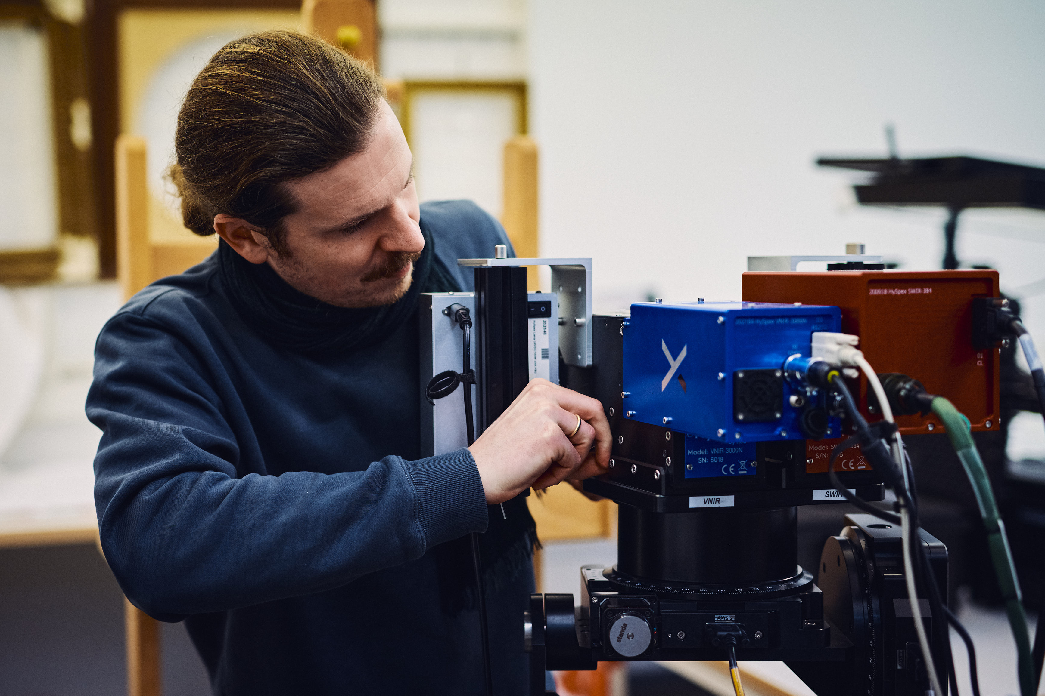
With the help of two cameras that together cover a very wide wavelength range of the electromagnetic spectrum, Simon Mindermann examines fragments of Willi Baumeister's works. Photo: Elia Schmid
This data can be used to distinguish between different materials and visualize their distribution on the surface of a painting (thereby creating a mapping).
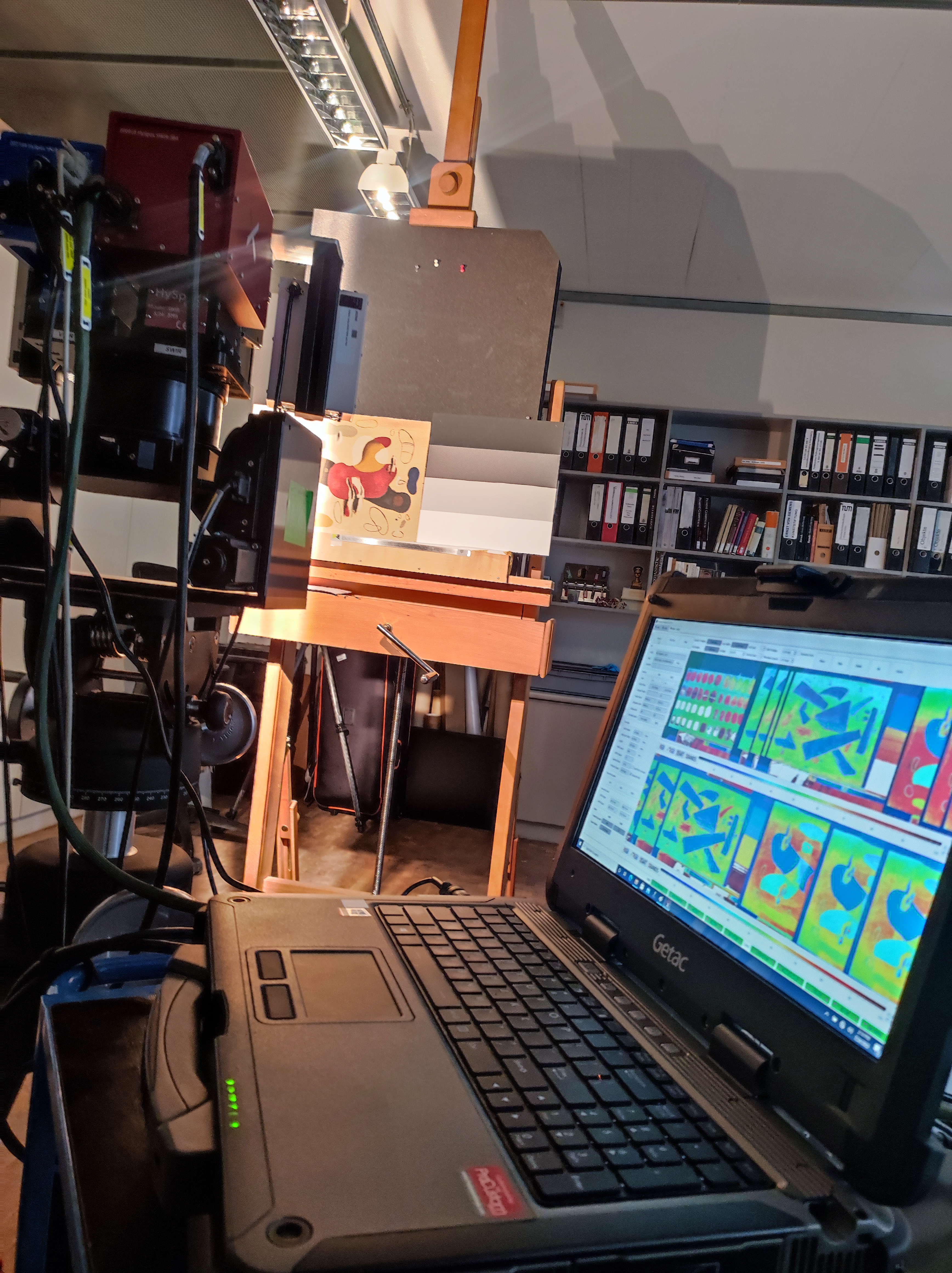
The data collected by the cameras is visualized and evaluated on a computer.
Photo: Katja Lorenz
HSI is already being used successfully to identify pigments and dyes in the visible and near-infrared range (VNIR, 400–1000 nm), but the investigation of binding media in the short-wave infrared range (SWIR, 1000–2500 nm), while possible, has only been tested in a few cultural heritage studies to date.
As part of her master's thesis, Katja Lorenz investigated the extent to which HSI is suitable for distinguishing and identifying typical binding media in Willi Baumeister's paintings, and she analyzed the possibilities and limitations of this method for art technology.
(Results from Katja Lorenz)
Non-destructive analysis methods have the great advantage of providing an initial, comprehensive overview of the distribution of materials on the painting surface without the need to take samples.
However, they mainly show what is visible on the surface. Normally, however, several layers of different materials lie on top of each other at a single measuring point. When radiation penetrates deeper layers, the signals from these different layers can overlap in the resulting spectrum. It then becomes unclear which material is actually contained in which layer. To allocate individual materials to particular layers within the layer structure of a painting, other – micro-invasive – methods are required.
With the help of non-destructive analysis methods, it is possible to make an initial classification of the existing materials into material groups such as proteins. However, more precise identification – that is, determining the exact type of protein that was used – usually requires further laboratory-based analyses. In addition, it is primarily the main components in a layer that can be detected by non-destructive methods. Minor components that occur in smaller quantities can easily be overlooked or are undetectable, because they are below the detection limit.
This leaves a number of questions unanswered regarding the layer structure and exact material composition, which will need to be clarified using micro-invasive, laboratory-based analysis methodsThese samples can then be examined in detail in the laboratory using high-resolution methods.
Although taking micro-samples for subsequent analyses is part of our research process and serves to gain knowledge, the decision is often difficult when it comes to original paintings, as it always involves some destruction – albeit very minor – of the original.
It is therefore fortunate for us that Baumeister discarded many paintings, but did not ultimately destroy them. These discarded paintings allow us to learn a great deal about the materiality of his artworks without having to sample the originals themselves.
When taking samples, we distinguish between two types: powder samples, which are taken by scraping with a fine scalpel, and samples of the entire layer package, which are then embedded in a hardening plastic and thus represent a cross-section of all layers of the painting (so-called cross-sections). The locations of the sampling sites on the paintings are measured very precisely and documented photographically. The amount of sample required is based on the so-called detection limit of the analysis techniques to be used: Each technique requires a minimum amount of material in order to achieve a meaningful result. The techniques used are very sensitive, which means that only very small samples are needed. One can clearly see in the two microscope images how small the sampling sites are.

Microscope image before sampling.
Photo: Ulrike Palm

Microscope image after sampling.
Photo: Ulrike Palm
Sampling site in the black paint layer of the discarded painting V_005. Top: before sampling, bottom: after sampling. The white frame marks the location of the scraping sample, which measures less than half a square millimeter. It is barely visible to the naked eye.
A particular focus of the project is the precise identification of the binding media used by Baumeister in his paintings.

The samples are found between two glass slides when they arrive at the laboratory.
Photo: Elia Schmid
The samples taken for this purpose are so small that they be seen and prepared for the subsequent analyses only under a microscope.

The samples are checked under the microscope before they are analyzed.
Photo: Elia Schmid
To determine the binding media, powder samples are examined using pyrolysis gas chromatography–mass spectrometry (pyrolysis GC–MS).
In this analytical technique, the sample is first heated to decompose it and convert it into volatile degradation products (via pyrolysis). The individual components are then separated using gas chromatography (GC) and subsequently identified with a mass spectrometer (MS).

Photo: Elia Schmid
Only a few laboratories in Germany specialize in the analysis of aged painting samples using pyrolysis GC–MS. This requires years of experience and considerable skill in handling the tiny samples.

Photo: Elia Schmid
The samples are first placed under a microscope into tiny metal sample containers measuring just a few millimeters in height. A precision scale is used to check that the minimum quantity of material required for analysis has been obtained.
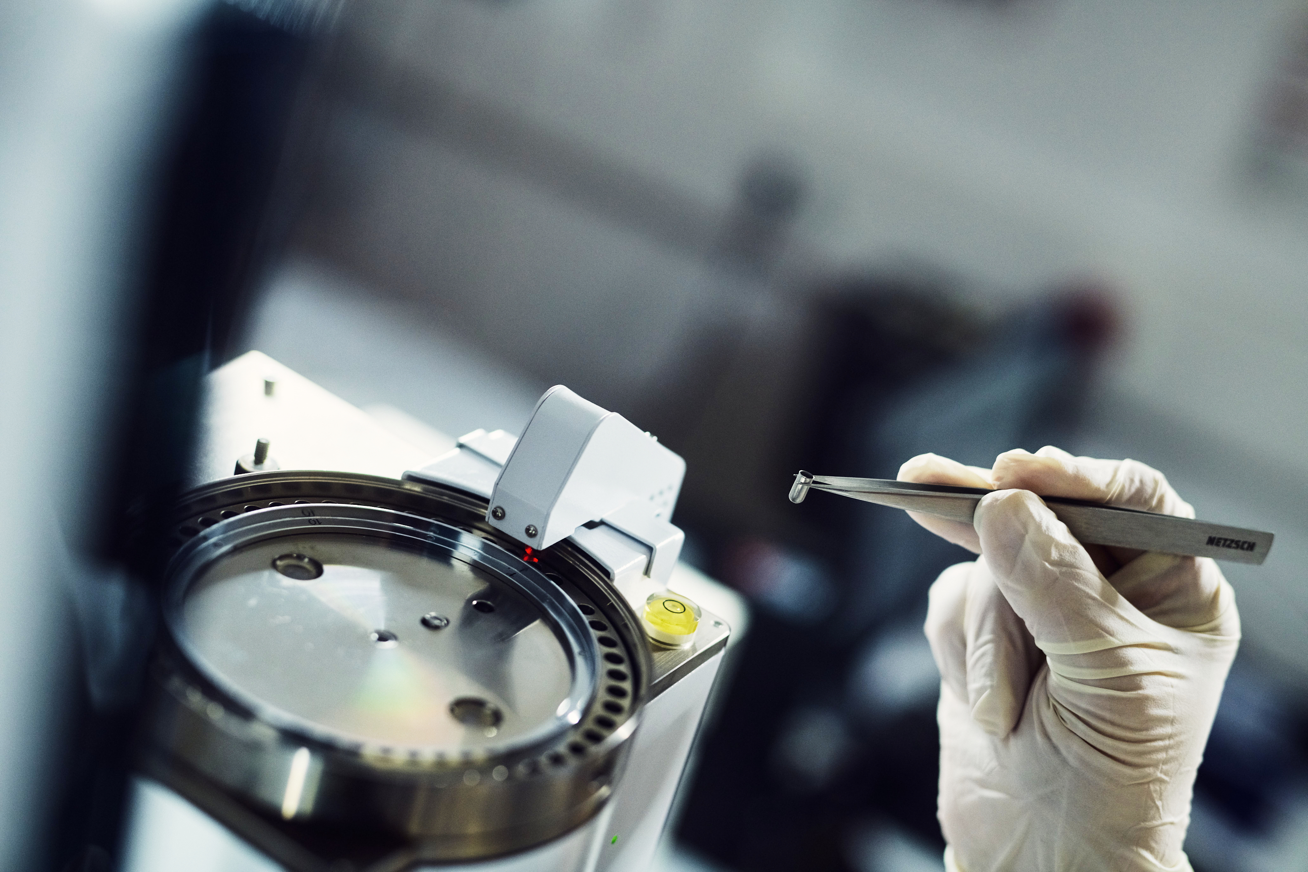
Photo: Elia Schmid
The containers are then placed in the sample chambers of the gas chromatograph.

With this analysis method, too, interpreting the results and comparing them with reference materials is very time-consuming. Photo: Elia Schmid
Once the analysis is complete, a chromatogram is produced. The individual peaks are characteristic of different substances and they aid in the identification of the individual substances in the sample.
The particular challenge in our field of research is that we are dealing with aged materials. Reference spectra of fresh samples are not necessarily comparable with those from aged samples. Alternatively, aged samples with a known composition can be measured. These can be found, for example, in collections of historical material samples. A high-quality interpretation of the results requires a lot of experience and a very good reference database that also includes reference samples whose composition has changed over time.

Chromatogram of a sample by Willi Baumeister
© Doerner Institute and Deutsches Museum, Munich.
In the chromatogram of a sample from Willi Baumeister, the individual peaks were assigned to different substances during evaluation: A synthetic resin (cyclohexanone) was identified, as well as several FC16, C18) from oils, and many different historical plasticizers.
Further investigations are also being carried out on the cross-sections: On the one hand, they can be examined under an optical microscope both in visible light and under UV radiation, thus revealing different layers – both transparent and pigmented layers. In this way, the stereomicroscopic observations of the layer structure can often be further refined and supplemented.
In this sample from a red paint layer of a discarded painting, the layer structure can be seen under the light microscope: a red paint layer lies on top of a light-colored layer – the ground layer. Under UV radiation, another very thin, light blue fluorescent layer, which was invisible in visible light, becomes visible on the surface of the red paint layer.
The scale also illustrates once again how small the sample is: 100 micrometers are equivalent to 0.1 mm.

Cross-section from a discarded painting viewed in visible light under a light microscope. Photo: Stephanie Dietz

The same cross-section under a light microscope under UV radiation. Photo: Stephanie Dietz
Using additional analytical methods, both the colorants (pigments) and the binding media in the individual layers can be determined by examining the cross-sections. Scanning electron microscopy with energy-dispersive X-ray spectroscopy (SEM-EDX) is used to determine the inorganic colorants, while a RAMAN spectrometer is used for the organic colorants. The distribution of the binding media in the layer structure can be examined using Fourier transform infrared spectroscopy with a focal plane array detector (FPA FT-IR imaging).
Each of the non-destructive and micro-invasive techniques described here has its particular strengths and weaknesses. Only when combined do they provide the most complete picture possible of the actual materiality of a painting. The aim is to obtain accurate and comprehensive information by skillfully combining them, while at the same time taking the fewest and smallest samples possible.
In this way, we are gradually learning more about the materials used by Baumeister between 1930 and 1955; step by step the picture is becoming more complete. The investigations of the paintings provide important pieces of the puzzle, along with the details in letters or invoices for painting materials. Ultimately, all the results must be compared and discussed by the research team.
However, the question then often arises as to the precise origin of the materials detected in the analyses of the paintings: Were certain substances such as synthetic resins components in artists' paints or industrial lacquers provided by Kurt Herberts? Comparative studies of historical tube paints, varnish bottles, and similar items from the artist's estate provide important clues. Source research in the archives of artists' paint manufacturers can also provide information about the composition of individual products that Baumeister purchased. This is the subject of Saskia Link's master's thesis (results).
In order to assess whether Baumeister's experiments in painting technique were unusual or phenomenon typical of the era, in which other German artists were involved, further sources must be evaluated. Besides present-day specialist literature, this includes contemporary how-to books on painting technique, such as the influential and widely read books by Max Doerner or Kurt Wehlte, and historical painting technique journals.
In the end, many questions have been answered – and at the same time, many new questions have arisen, which can be pursued in the future.